
Relevance
How does the human brain decide what information to attend to and what information to ignore? Relevance detection refers to neural processes determining the salience of a stimulus.
General Considerations
According to the Appraisal Theory of Emotion, relevance detection is mainly modulated by three variables:
i) Novelty (in terms of familiarity and/or predictability)
ii) Pleasantness (in terms of neutral, positive or negative valence)
ii) Goal / Need Relevance (in terms of social versus nonsocial or communicative relevance, etc.)
In a number of research projects, Dr Vrticka is trying to disentangle the effects of these three variables on brain activity, both on a temporal ([i]EEG) as well as a functional anatomical (fMRI) level. In doing so, a particular focus is directed on how the human brain represents an additional relevance category, namely:
iv) Social (versus Nonsocial) Content
Although social information is known to be of high intrinsic relevance to the human organism, and previous fMRI investigations have shown that it likely is processed as a unique stimulus dimension, it remains largely unknown how relevance detection in terms of novelty, pleasantness, and goal/need relevance may interact with the processing of social relevance. One region of interest for fMRI analyses is the amygdala (see references by Sander, Ousdal, as well as Pessoa and Adolphs), but other brain areas are considered as well. Dr Vrticka is also interested in individual differences that may influence relevance detection.
Please see below for a more thorough description of the individual studies related to relevance detection, which are:
1) Social versus emotional relevance – fMRI in healthy adults
2) Social versus emotional relevance – EEG in healthy adults
3) Social versus emotional relevance – combined EEG & fMRI in healthy adults
4) Social versus emotional relevance – iEEG in Epilepsy Patients
5) Social versus emotional relevance – 22q11DS patients versus controls
6) Social versus emotional relevance versus novelty – fMRI in healthy adults
1) Social Versus Emotional Relevance – fMRI in Healthy Adults
In one study published in Frontiers in Human Neuroscience (2013), we investigated the neural substrates of social versus emotional relevance processing. To do so, we presented images depicting complex visual scenes that could either have a social or nonsocial content, and be of either positive, negative or “neutral” valence to healthy adult participants.
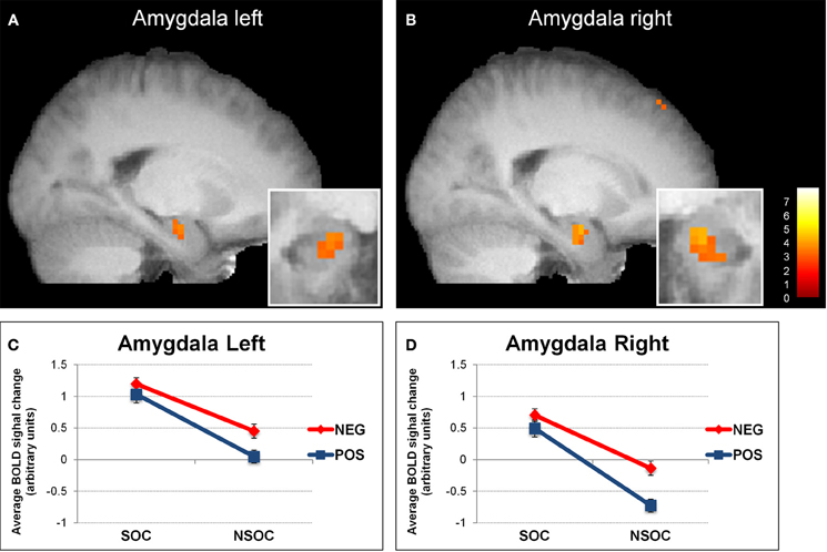
We found that in several brain areas, comprising the bilateral amygdala, fusiform gyrus, anterior superior temporal gyrus and medial orbitofrontal cortex, brain activity reflected interactive processing of social content and emotional valence.
Firstly, activity was always higher to emotional versus neutral images. Second, activity was always higher to social versus nonsocial images. And third, while activity to social images did not significantly differ as a function of positive versus negative valence, it was higher for negative versus positive valence for nonsocial images (see Figure on the right illustrating such pattern in bilateral amygdala).
Our findings suggest that the human brain, or at least the regions mentioned above, attribute(s) stronger relevance to social (versus nonsocial) information, regardless of valence. This reflects the notion that social information is highly salient for human behavior per se. In turn, probably because of the higher biological salience in terms of survival, nonsocial negative (versus positive) information is preferentially processed, allowing for a fast detection of potential threats.

Interestingly, the above social by emotional relevance interaction in the right amygdala was modulated by the participants’ trait anxiety: the higher the participants’ trait anxiety, the weaker the interaction became (i.e., for high trait anxious participants, there was only a main effect of social and emotional relevance, but no longer an interaction). This effect was driven by a selective decrease of activity for the social positive condition with increasing trait anxiety scores. A putative explanation could be stronger habituation due to subjectively perceived lower relevance of social positive information for high trait anxious people.
See also this blog post for another summary of obtained results.
2) Social Versus Emotional Relevance – EEG in Healthy Adults
To build upon the above published fMRI data, Annekathrin Schacht (University of Göttingen, Germany) and Dr Vrticka took the same complex visual scenes as used in the fMRI study and embedded them into an EEG task to investigate the more precise temporal unfolding of social and emotional relevance processing. Our main questions were: (i) which of these two stimulus dimensions is processed first – i.e., which relevance check occurrs first -, and (ii) when in time social and emotional relevance processing interacts as suggested by our fMRI data – as opposed to be processed separately and/or additively.

Interestingly, we found that social and emotional relevance was processed interactively already at very early stages of visual processing, namely at around 80-120 ms after stimulus onset during the P1 – especially for social positive versus nonsocial positive content. Such interactive processing continued around 200-320 ms during the EPN with a slightly different pattern. Furthermore, the interaction pattern observed with EEG strongly resembled the interaction pattern detected with fMRI (especially the pattern between 200-320 ms during the EPN), and source localization of the ERP components of the social > nonsocial effect revealed strongly overlapping neural substrates. The corresponding paper is published in CABN (open access).
See also this blog post for another summary of obtained results.
3) Social Versus Emotional Relevance – Combined EEG & fMRI in Healthy Adults
As a logical next step – having looked at social and emotional relevance processing in fMRI and EEG separately – Annekathrin Schacht & Simon Weber (University of Göttingen, Germany) and Dr Vrticka ran a follow up study during which EEG and fMRI data using the same stimuli was acquired from the same participants. In so doing, we will be able to combine the fMRI and EEG data to obtain an even more elaborate spatiotemporal pattern of social and emotional relevance processing. The data is currently being written up for publication.
4) Social Versus Emotional Relevance – iEEG in Epilepsy Patients
Having assessed social content and emotional valence processing with skalp EEG so far, we were interested in obtaing more precise anatomical localization of observed ERP patterns. To do so, Pascal Vrticka and Annekathrin Schacht (University of Göttingen, Germany) teamed up with Petr Marusič, Jiří Hammer & Alena Javůrková from the Department of Neurology, Motol University Hospital & Faculty of Medicine, Charles University, Praha (Czech Republic), where epilepsy patients are implanted with intracranial EEG (iEEG) electrodes. Once more, we are using the same complex visual scenes to determine when and particularly where social content and emotional valence are processed within the human brain. The study is currently underway.
5) Social Versus Emotional Relevance – fMRI in 22q11DS Patients Versus Controls
As part of a collaboration with the University of Geneva, we again used the same social versus nonsocial positive versus negative images to look at the neural substrates of social versus emotional relevance detection in 22q11DS patients versus healthy controls. The 22q11.2 deletion syndrome is a neurogenetic disorder associated with elevated risk for schizophrenia and high rates of distinct positive and negative symptoms. Social impairments were also described in this population. However, the neural correlates underlying these impairments are largely unknown.
The observed results indicate atypical neural activation during social perception (social versus nonsocial images) in 22q11DS patients that appear to be independent of valence processing (no differences between groups).
The corresponding paper is published open access online and can be downloaded here.
6) Social Versus Emotional Relevance versus Novelty – fMRI in Healthy Adults
In another fMRI study published in Emotion (2013), we examined brain responses to dynamic emotional facial expressions and comparable nonsocial stimuli (“lottery game”) as a function of stimulus novelty and valence. Novelty was conveyed by the presence versus absence of surprise, while valence was reflected by positive versus negative versus neutral stimulus content. We were especially interested in how the amygdala processes such different kinds of stimulus relevance.
Our findings revealed an intriguing activation pattern for both social and nonsocial stimulus processing. The amygdala did not seem to differentiate between valence and novelty as such, but was more strongly activated to dynamic information containing positive versus negative surprise. According to the available literature, and particularly a previous study by Yacubian et al. (2006), such findings suggest that the human amygdala – at least under conditions of uncertainty involving surprise conveyed by dynamic facial expressions – (i) encodes a negative expected value (anticipation of a negative outcome), and subsequently (ii) updates such expectation by means of a negative expected value prediction error if a positive outcome occurs instead.
Our data propose that relevance processing in the human amygdala may not only be modulated by the static versus dynamic nature of stimulus presentation, but also depend on the degree of uncertainty / predictability of information.
For more detailed information regarding the above research on relevance detection, please refer to the Publications page or contact Dr Vrticka with any questions.